2
1.8yr
A video of a "giant" single-celled organism eating a rotifer!
Breakthrough here is the ability to image embryos comprised of living cells as opposed to post-mortem embryos.
original doi: https://doi.org/10.1016/j.cell.2023.06.003
Next project. :)
I've gotten all the parts printed and collected but haven't set it up yet. Excited to see how well it does.

Tender-handed, stroke a nettle, ... read full post
This summarizes a paper in which researchers created a microscope probe using a single, 110 µm diameter optical fiber. An interesting technique from this that I had not seen before, was that the end of the optical fiber is angled and coated with aluminum to create a mirror looking perpendicular to the length of the fiber. This enables researchers to image living cells less intrusively. ... read full post
Tarantula (Acanthoscurria geniculata) embryo showing expression of genes that pattern the legs and body wall. Right side has curved abdomen, 4 walking legs, and 1 pedipalp (head accidentally removed). Left side has curved abdomen and 1 walking leg (the rest accidentally removed).
The pink/white stripes are where the gene "odd-skipped" is expressed, which marks where the leg joints will form.
Error message is SyntaxError: Unexpected token 'R', "Request er"... is not valid JSON
Are image uploads not allowed here? Only URLs? Thanks! Hoping this community grows and flourishes! :)
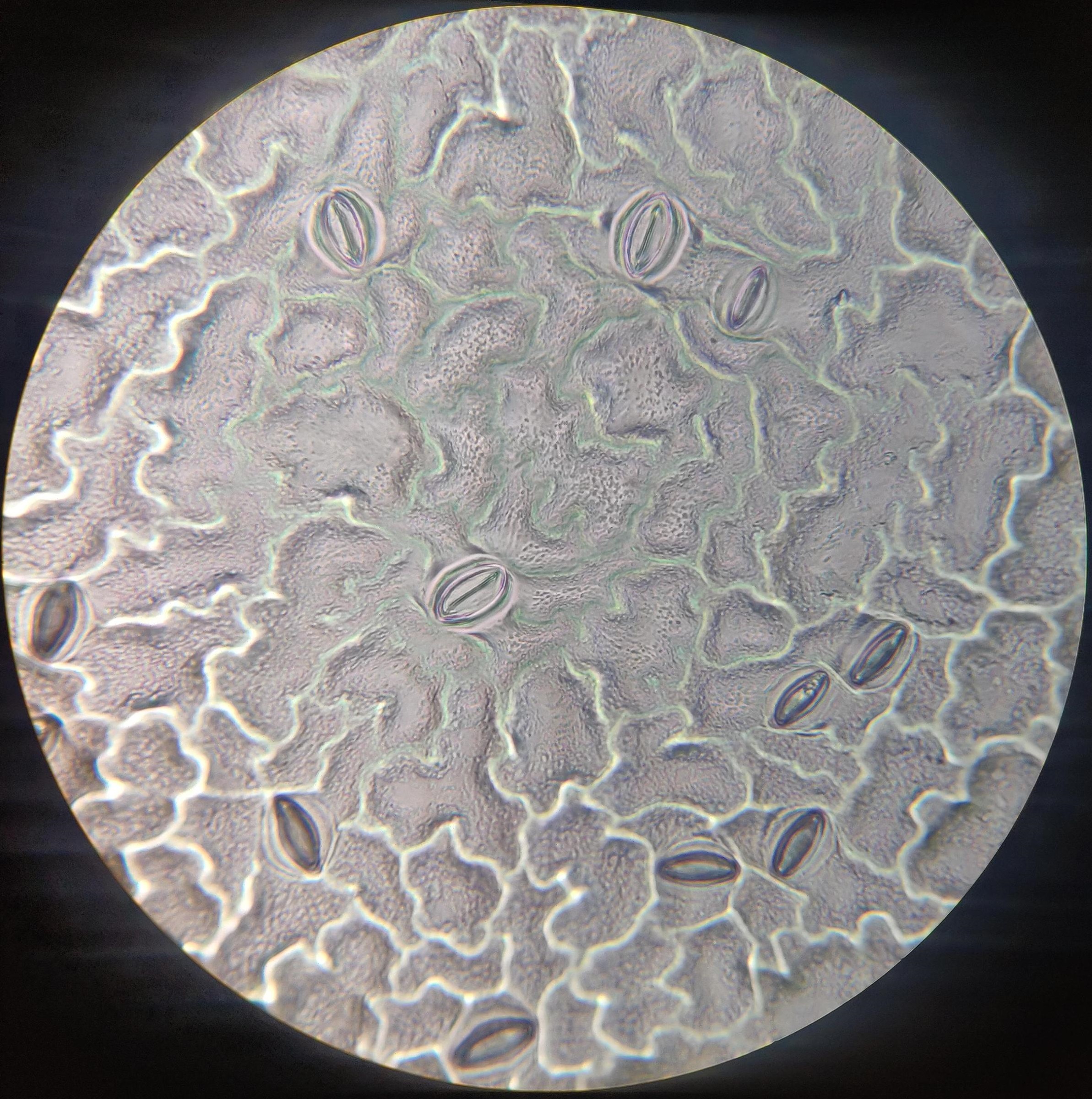
The school year just ended but I wanted to show off what my class was able to accomplish with some cheap high school microscopes. The images below are under-leaf impressions of diocot and monocot plants which show the stomata used to control water evaporation. To avoid the preparation of slicing leaves themselves we painted a thin layer of nail polish on the underside. Peeling this dry layer off results in a mold of the leaf itself which can then be attached to a blank slide with cellophane tape. ... read full post

I have to admit, not my most successful attempt at the "apply violence until the die is visible" method of decapping ever. The silver stuff to the right is the broken off silicon.
I put a drop of liquid from my lotus flower's vase under the microscope and noticed a lot of bdelloid rotifers, so I decided to record one at 400x magnification. ... read full post